ESPCI web site

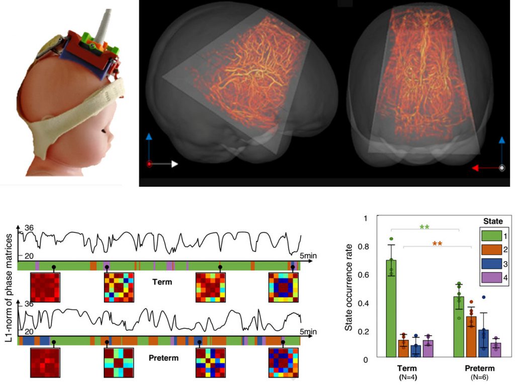
Functional ultrasound imaging reveals the dynamics of brain states in preterm babies
Our laboratory Physics for Medicine Paris (Inserm/ESPCI Paris-PSL/CNRS), in collaboration with Robert Debré Hospital (AP-HP/Inserm) is developing functional ultrasound imaging to monitor the brain activity of neonates in a way that existing neuroimaging modalities do not achieve. Our latest study, published this month in Nature Communications, marks another leap forward by demonstrating that functional ultrasound allows to monitor and analyze functional connectivity patterns between different brain regions and their evolution in time in neonates. This new tool could be used for an early prediction of brain disorders and neurological outcomes of premature birth.
Prematurity can affect the development of the neonate’s brain and result in neurological impairments of various degrees of severity, some having life-long consequences. Examples of pathologies include autism spectrum disorders, intellectual deficiency, epilepsy, attention deficit and hyperactivity disorders, etc. A major challenge in neonatal medicine is to detect and identify the neurodevelopmental trajectory of neonates at the earliest stage after birth. Monitoring their cerebral functions is however difficult with existing neuroimaging tools.
Functional MRI (magnetic resonance imaging) is the gold-standard for neuroimaging, but it is hardly applicable for a longitudinal monitoring of vulnerable subjects such as preterm neonates, given its cost, its cumbersomeness and sensitivity to motion artifacts. EEG (electro-encephalography) and NIRS (near-infrared spectroscopy) can be used at the neonate’s bedside, but have a limited spatial resolution and imaging depth. This is where functional ultrasound (fUS) comes in: through its unrivalled sensitivity and great portability, functional ultrasound imaging will potentially become a powerful tool to monitor the babies’ brain functions. The examination is indeed performed at the baby’s bedside by placing an echographic probe on the neonate’s head.
In 2017, we made the first demonstration of acquiring functional ultrasound data in human neonates. More recently, we have been working on analysis methods to extract from these data functional connectivity information. Functional connectivity is the existence of functional relationships – in particular synchronization of the activity – between different regions of the brain. Alterations in the functional connectivity can be a marker of cerebral dysfunctions. In the present study, we recorded the brain activity of neonates at rest, and not only did we extract functional connectivity patterns, but we also studied their evolution in time and the occurrence rate of each pattern (see figure below). We derived three methods to assess the functional connectivity, each exploiting different characteristics of the data and providing complementary information. Dynamic functional connectivity is a topic of interest in neuroimaging and has been studied in adult subjects using MRI, but little data has been obtained on neonates so far. The first results obtained using functional ultrasound in a small cohort suggest a significant difference between term and preterm babies.
This study takes functional ultrasound one step further towards clinical applications, by defining advanced analysis methods to provide a fine and robust assessment of both static and dynamic functional connectivity. We believe that the robustness and ease of use of functional ultrasound imaging will greatly facilitate clinical investigations, and ultimately provide a new and powerful tool in neonatal care.
Full publication:
Baranger J, Demené C, Frerot A, Faure F, Delanoë C, Serroune H, Houdouin H, Mairesse J, Biran V, Baud O, Tanter M. Bedside functional monitoring of the dynamic brain connectivity un human neonates. Nature Communications 2021