 ESPCI web site

Ultrafast ultrasound imaging of the spinal cord in front page of Pain journal
Pain modulation is a mechanism which starts in the spinal cord: pain signals are processed by the spinal cord before being sent to the brain. Studying the spinal cord physiology and its alterations is therefore essential to understand how pain can be modulated either through natural events (e.g. during stress or physical effort) or using pharmaceutical drugs (e.g. morphine). We recently combined two innovative ultrasound imaging techniques – functional ultrasound imaging (fUS) and ultrasound localization microscopy (ULM) – to map in vivo for the first time the spinal cord vasculature up to microscopic resolution and its neurofunctional response to innocuous stimuli. The study has just been published in front cover of Pain journal and chosen as the editor’s highlight.
The role of the spinal cord in pain processing has been extensively studied using electrophysiology (in vivo) or histochemistry staining (ex vivo). However, these methods only provide a local and static measurement of the neuronal activity. Our innovative ultrasound-based imaging methods cover entire cross-sections of the spinal cord, are applicable in vivo in rats, and provide a highly sensitive and highly resolved (in space and time) assessment of the spinal cord hemodynamics.
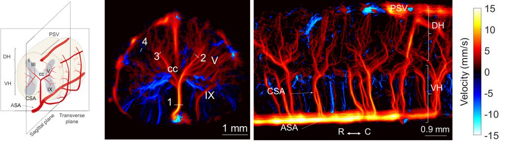
- Ultrasound localization microscopy (ULM) reveals the vascular network of the spinal cord, in transverse and sagittal planes, with a 10-micron spatial resolution.
- Functional ultrasound (fUS) allows us to investigate the neurovascular response of different areas of spinal cord to peripheral stimuli (applied to the hind paw of anesthetized rats). We demonstrate that the spinal cord response depends upon the type of activated nerve fibers and involves the glutamatergic NMDA receptor. In a pathology model of inflammation, we also observed an exacerbated response to innocuous stimuli.
The high sensitivity of functional ultrasound imaging enabled us to map and perform for the first time a fine analysis of the hemodynamics in different areas of the spinal cord in response to pain conditions. Our study sets the foundations for deploying functional ultrasound imaging as a powerful tool to develop analgesic drugs. Pain journal acknowledged the importance of our study by selecting it as the front cover of this month’s issue and as the Editor’s highlight of the journal.
Watch the video abstract here.

Full-text publication: Claron J, Hingot V, Rivals I, Rahal L, Couture O, Deffieux T, Tanter M, Pezet S. Large-scale functional ultrasound imaging of the spinal cord reveals in-depth spatiotemporal responses of spinal nociceptive circuits in both normal and inflammatory states. PAIN 2021;162:1047–59. https://doi.org/10.1097/j.pain.0000000000002078.
Cover designed by Alexandre Dizeux


