Quantification and ultrasound biomarkers
SCIENTIFIC LEADER
Clément Papadacci
KEYWORDS
-
Elastography and tissular biomechanics
-
Fiber tractography
-
Ultrasensitive Doppler imaging
-
Quantitative diagnosis of steatosis
-
Ultrasound super-localization microscopy
-
3D ultrafast ultrasound imaging
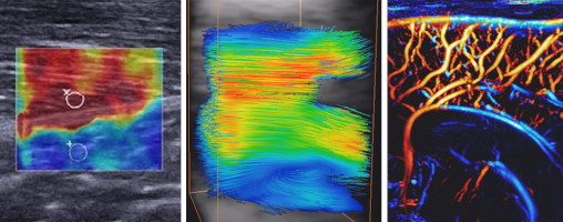
We are developing new and non-invasive methods to better characterize the properties of tissues and blood flow, in order to define efficient biomarkers for diagnostic purposes. The clinical feasibility of these methods has been established in various applications in cardiology, neurosciences or oncology. For instance:
- Shear wave elastography maps the mechanical properties of tissues and is applied to assessing the contractility of muscles (in particular the cardiac muscle), or providing an early diagnosis of cancer (through non-invasive examinations that have already halved the number of biopsies in breast cancer diagnosis).
- Blood flow can be quantified over large fields of view, down to the smallest vessels: the ultrasensitive Doppler imaging method allows imaging the perfusion of organs such as the brain, the kidney or the liver.
- The fat content in tissues can be measured with ultrasound for a quantitative diagnosis of hepatic steatosis.
- Combining ultrafast ultrasound imaging and contrast agents, ultrasound super-localization microscopy multiplies the spatial resolution of vascular maps, allowing to monitor tumor angiogenesis (formation of microvessels during tumor growth) or to detect fine vascular lesions.

Main publications
- Tanter M et al. Quantitative Assessment of Breast Lesion Viscoelasticity: Initial Clinical Results Using Supersonic Shear Imaging. Ultrasound Med Biol (2008)
- Papadacci C et al. Imaging the dynamics of cardiac fiber orientation in vivo using 3D Ultrasound Backscatter Tensor Imaging. Sci Rep (2017)
- Demené C et al. 4D microvascular imaging based on ultrafast Doppler tomography. Neuroimage (2016)
- Imbault M et al. Robust sound speed estimation for ultrasound-based hepatic steatosis assessment. Phys Med Biol (2017)
- Errico C et al. Ultrafast ultrasound localization microscopy for deep super-resolution vascular imaging. Nature (2015)