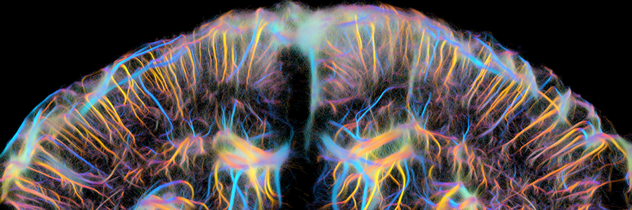
Non-invasive characterization of pericyte dysfunction in mouse brain using functional ultrasound localization microscopy: a major advance for diagnosing vascular and neurovascular alterations
Coordinated by Physics for Medicine Paris in collaboration with Leiden University Medical Center, our study published in Nature Biomedical Engineering demonstrates for the first time that it is possible to image non-invasively the cerebrovascular alterations due to pericyte dysfunction in the mouse brain. Pericytes are small contractile cells located on the walls of capillaries that help regulate cerebral blood flow. These cells play a key role in the early stages of many cerebrovascular diseases and are therefore a promising target for developing new treatments.
However, the mechanisms linking pericyte dysfunction to changes in cerebral vascularization remain only partially understood, and to date, no reliable non-invasive method exists to identify them and to monitor the efficacy of therapeutic treatments.
The power of ultrasound localization microscopy
In this study funded by the EIC Health Pathfinder project MICROVASC, our team used functional ultrasound localization microscopy (fULM), a super-resolution imaging technology developed in 2022. It enables whole-brain visualization with unmatched precision, without surgery.
Using a rare genetic disease model — hereditary hemorrhagic telangiectasia — we observed typical abnormalities of pericyte dysfunction: capillaries with irregular shapes and increased diameters, reduced velocity in arterioles, and impaired neurovascular coupling. These non-invasive observations constitute promising biomarkers for monitoring pericyte dysfunction at the whole-brain scale.
Finally, ultrasound imaging revealed that treatment of mice with molecule (C381) activating cellular TGF-β signaling restores the structure of cerebral arteries and their hemodynamic responses.
Conducted in collaboration with the Leiden University Medical Center (Franck Lebrin’s team), our study shows for the first time that it is possible to fully non-invasively image vascular and neurovascular alterations of pericytes and small brain vessels, as well as their functional recovery under various therapeutic treatments.
With the development of super-resolution ultrasound imaging in humans, this work paves the way for a better understanding of small vessel diseases and the study of pericyte-targeting treatments in patients.
📑Learn more about our fULM technology
🇫🇷 For more details, see the French press release | La microscopie ultrasonore fonctionnelle (fULM) : une technique révolutionnaire pour détecter et diagnostiquer les maladies des plus petits vaisseaux cérébraux


