 ESPCI web site

3D ultrasound microscopy of the brain on the front cover of eBioMedicine
Our researchers obtained fantastic images of the brain vasculature using 3D ultrasound localization microscopy (3D ULM). The study is on the front cover of eBioMedicine on May 2022 issue, and closely follows our previous publication on the imaging of the coronary microcirculation.
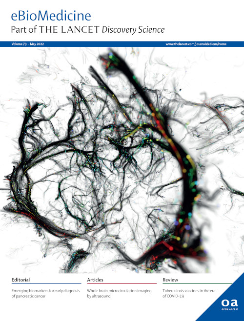
In the recent years, researchers of Physics for Medicine Paris (Inserm, ESPCI Paris-PSL, CNRS) have developed major advances in the field of vascular imaging, with the advent of ultrasensitive Doppler imaging in 2008, and then ultrasound localization microscopy (ULM) in 2015. They have now succeeded in deploying ultrasound microscopy in 3D for transcranial whole-brain imaging.
The results show the vascular network of the entire mouse brain, imaged through the skull, with a micron scale resolution. To obtain such a fine spatial resolution, biocompatible microbubbles are injected in the blood circulation, and tracked at a high frame rate (several thousands of volumes per second) using 3D ultrafast ultrasound imaging. The collected data can then be analyzed to retrieve detailed information on the brain local hemodynamics: the diameter and blood flow velocity can be quantified in each vessel.
The cerebral vascular network is essentiel to support and regulate the brain functions. Stroke and aneurysms are examples of cerebrovascular dysfunctions, but vascular alterations are also now known to play a role in neurodegenerative diseases. Observing and characterizing the blood flow dynamics across several spatial scales opens new perspectives for detecting and diagnosing the disease at an early stage. The technology is licensed to the company Iconeus, which already commercializes ultrasound neuroimagers for preclinical brain research. On the clinical side, ultrasound microscopy of the brain was achieved in 2D in human patients in 2021. The demonstration of transcranial 3D ULM in rodents is a further technological step towards a clinical deployment of whole-brain ultrasound microangiography.
Full publication: Demeulenaere O*, Bertolo A*, Pezet S, Ialy-Radio N, Osmanski B, Papadacci C, Tanter M, Deffieux T**, Pernot M**. In vivo whole brain microvascular imaging in mice using transcranial 3D Ultrasound Localization Microscopy 2022;79:12. https://doi.org/10.1016/j.ebiom.2022.103995
Corresponding author: mathieu.pernot@espci.fr
Video credits: Alexandre Dizeux / Physics for Medicine Paris


