 ESPCI web site

3D ultrafast echocardiography: full characterization of cardiac functions within a heartbeat
Imaging the heart in three dimensions at >1000 volumes per second allows to assess a whole set of parameters of the heart functions within a single cardiac cycle. Two of our recent studies, performed on healthy volunteers, are in the news: one is featured in the highlights of the EuroEcho conference, while the other has just been published in IEEE Transactions on Medical Imaging.
The heart works around the clock through a repeated sequence of contractions and relaxations, ejecting and pumping blood to supply all the body with oxygen. Dysfunctions in this cardiac cycle lead to heart failure, a common pathology with various degrees of severity. This constantly moving organ is a challenge for biomedical imaging. Echocardiography (i.e. ultrasound imaging of the heart) is the commonest imaging modality used in hospitals. In our lab, we have developed ultrafast echocardiography, to capture all the mechanical events and blood flows occurring within a single cardiac cycle by acquiring thousands of frames per second.
The first ultrafast ultrasound scanners were however limited to two-dimensional imaging, providing only cross-sectional views of the heart that fail to capture properly out-of-planes blood flows and tissue motions. 2D acquisitions are also variable as they highly depend on the positioning of the ultrasound probe. In 2014, we built the very first 3D ultrafast ultrasound scanner, capable of acquiring thousands of volumes per second. Volumetric imaging is essential to characterize the heart’s three-dimensional motions in a quantitative and robust manner.
In 2019, we applied 3D ultrafast echocardiography to quantify Doppler indices within a single cardiac cycle. Doppler indices are an estimate of the blood volumes pumped in and ejected from the heart during the cardiac cycle. They are routinely assessed using conventional echocardiography with however a high variability due to the 2D limitation and low frame rate. Our work using 3D ultrafast echocardiography was in the spotlight of the 2019 EuroEcho conference in Vienna, Austria, where it has been selected amongst 1639 abstracts to enter the Young Investigator Award Competition and the conference’s Highlights.
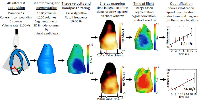
Last month, we published another study where 3D ultrafast echocardiography was derived to image the natural mechanical waves occurring in the heart as a result of the valves closure during the cardiac cycle. Tracking these waves provides information on the stiffness of the cardiac muscle, a key parameter that defines the heart ability to properly contract/relax.
These two studies demonstrate the feasibility of 3D ultrafast echocardiography on human healthy volunteers, with the development of new imaging and processing methods to quantify the heart functions. Performing acquisitions within a single cardiac cycle It could transform the practice of cardiology by providing a robust and operator-independent tool for diagnosis of heart dysfunctions. The next steps will aim at refining the methods and apply them to patients to demonstrate their clinical value.
References:
- Papadacci C, Finel V, Villemain O, Tanter M, Pernot M. 4D ultrafast ultrasound imaging of naturally occurring shear waves in the human heart. IEEE Transactions on Medical Imaging 2020:1–1. https://doi.org/10.1109/TMI.2020.3020147.
- Papadacci C, Finel V, Villemain O, Goudot G, Provost J, Messas E, et al. 4D simultaneous tissue and blood flow Doppler imaging: revisiting cardiac Doppler index with single heart beat 4D ultrafast echocardiography. Physics in Medicine and Biology 2019;64:. https://doi.org/10.1088/1361-6560/ab1107.


